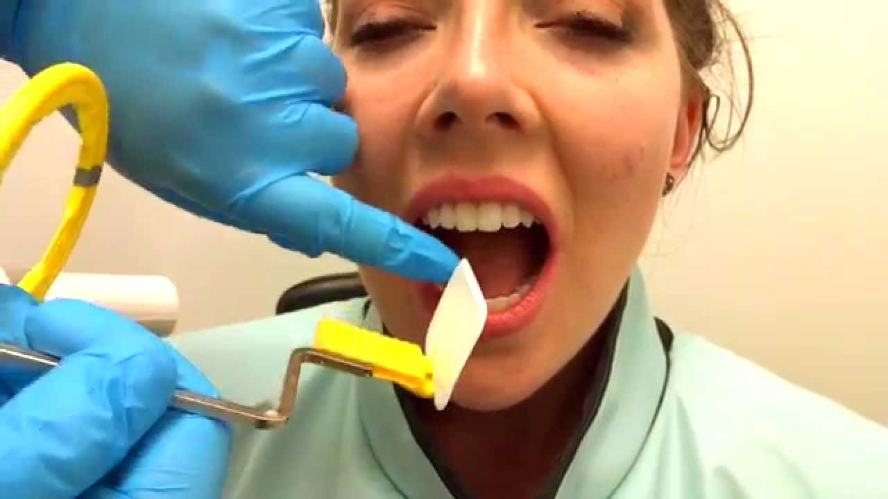
The film is placed parallel to the long axis. Demonstration on how to take periapical x-ray using bisecting angle technique.

Periapical Radiography Pocket Dentistry
Some further techniques to make this procedure more comfortable for the patient include.

. The patient was positioned upright with hisher mouth was opened as wide as possible to allow the X-ray beam to pass to the sensor unobstructed from the opposite side of the mouth. Fitzgerald called as paralleling or long cone technique. Bending the film produces an artifact on the image that may render the film unusable.
In order to minimize image distortion the. What are periapical radiographs used for. 3 What is an intraoral technique of exposing periapical images.
Periapical X-rays are used to detect any abnormalities of the root structure and surrounding bone structure. To take a periapical exposure the hygienist or x-ray technician places a small photosensitive imaging plate coated with phosphorus into a sterile wrapper and inserts it into the patients mouth just like a conventional X-ray film card. The X-ray tubehead is then aimed at right angles vertically and horizontally to both the tooth and the image.
Film must be positioned parallel to the long axis of the tooth. The image receptor is placed in a holder and positioned in the mouth parallel to the long axis of the tooth under. Preparing a Patient for the Paralleling Technique.
The dental assistant exposing a periapical film sensor using the paralleling technique should always start with the. The accelerated x-rays are decelerated by the target material resulting in bremsstrahlung. In an attempt to overcome the problems two techniques have been developed for periapical radiography.
The X-ray is taken and the exposed plate is then loaded into a scanner or processor which reads the image. When comparing the two periapical techniques the advantages of the bisecting angle technique are. Parallel technique The image receptor is placed in a holder and placed in the mouth parallel to the longitudinal axis of the tooth under investigation.
Ad Publishing Open Access Peer Reviewed Research related to the field of Scanning. Each periapical X-ray shows all teeth in one portion of either the upper or lower jaw. Periapical radiography is a commonly used intraoral imaging technique in radiology and may be a component of your radiologic examination.
The snap-a-ray is used. With the proposed software only a periapical X-ray is provided so the software cannot provide a diagnosis that. The film is placed parallel to the long axis of the tooth to be radiographed and the central beam of X-ray is directed at right angle to the film and the teeth.
Periapical lesions a c ommon dental disease are conventionally detected by dentists through radiog raphy. The bisected angle technique. Periapical radiographs provide important information about the teeth and surrounding bone.
4 Which technique is most often used when exposing a periapical image. Periapical film is held parallel to the long axis of the tooth using film-holding instruments. Most frequently used radiography is for the periapical which is performed by the bisecting Thus when considering the execution of the radiographic technique and the possibility of errors that occur during the exposure of X-ray image XR receptors it is important to identify those that occur more frequently.
The sensor was placed on the. Basically there are two techniques for taking periapical radiography. First to diagnosis periapical lesions a thorough clinical examination about dental caries tooth mobility vertical percussion sinus tract and X-ray is required.
For taking periapical radiographs on most adult patients you need size 2 film for posterior areas and size 1 or 2 for anterior areas. Periapical X-rays detect any unusual changes in the root and surrounding bone structures. Gently softening the edges of the film by rounding it very carefully between the fingers.
The technique of parallelism. The paralleling technique results in good quality x-rays with a minimum of distortion and is the most reliable technique for taking periapical x-rays. 6 Which exposure technique does the American Academy of Oral and Maxillofacial Radiology and the American Dental Education Association recommend.
Paralleling technique Bisecting angle technique Paralleling technique It is also called the extension cone paralleling technique right angle technique and long cone technique. However if the childsmouth is large enough to accommodate size 1 or 2 film and the child is cooperative use the larger size film. PARALLELING LONG-CONE PERIAPICAL EXPOSURE TECHNIQUES GENERAL A long cone is used to take x-rays with paralleling exposure techniques.
5 What is bisecting angle technique in dentistry. DENTAL X-RAYS X-rays are produced by boiling off electrons from a filament the cathodeand accelerating the el to the target at the anode. Submit Your Manuscript With Hindawi.
For this purpose a special technique of periapical radiography was developed by Gordon M. For children with small mouths you will need size 0 film. Join Leading Researchers in the Field.
Because the film is placed in the mouth at an angle to the long axis of the teeth. Size 2 Film is used for Anterior and Posterior X-rays when Bisecting. This must be done only slightly and the film must never be bent outright.
The central ray is directed to pass at a perpendicular angle to both the tooth and the film. The paralleling technique results in good quality x-rays with a minimum of distortion and is the most reliable technique for taking periapical x-rays. The extraoral periapical radiographic technique was performed for both maxillary and mandibular teeth using Newman and Friedman technique2.
The paralleling technique is considered to be the best way to take periapical X-rays and when used correctly it should produce reliable images with minimal distortion. By using a filmsensor holder with fixed image receptor and. The film is placed parallel to the long axis of the tooth in question and the central x-ray beam should be directed perpendicular to the long axis of the tooth.
This study aimed to assess the application of.
How To Take Periapical Radiographs Youtube
Periapical Radiography Pocket Dentistry

Periapical Radiography Pocket Dentistry

Periapical Radiography Pocket Dentistry

Periapical Radiography Pocket Dentistry

How Make Periapical X Ray

Periapical Radiography Pocket Dentistry

Periapical Radiography Pocket Dentistry
0 comments
Post a Comment